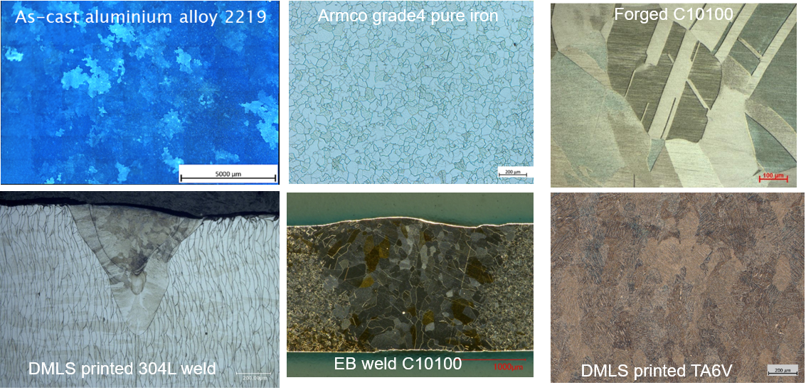
Optical microscopy is a widely used technique for analysing metallographic specimens. The typical magnification range for optical microscopes is from ×50 to ×1000. Optical microscopes use a number of different optical techniques to reveal specific microstructural features, including the following illumination techniques: bright field (BF), dark field (DF), polarized light (POL), oblique (stereo) and Differential Interference Contrast (DIC):
BF illumination is the most common illumination technique for metallographic analysis. The light path for BF illumination is from the source, through the objective, reflected off the surface, returning through the objective, and back to the eyepiece or camera.
DF illumination is a lesser known but powerful illumination technique. The light path for DF illumination is from the source, down the outside of the objective, reflected off the surface, returned through the objective and back to the eyepiece or camera.
DIC is a very useful illumination technique for providing enhanced specimen features. DIC uses a Normarski prism along with a polarizer in the 90° crossed positions. Essentially, two light beams are made to coincide at the focal plane of the objective, thus rendering height differences more visible as variations in colour.
Few examples of micrographs with different alloys, etching and illuminations methods are displayed below.
