The Focused Ion Beam in dual beam configuration with Scanning Electron Microscope (FIB-SEM) is the most advanced electron microscope apparatus within the section. This system combines the use of the electron beam for imaging and analysis with an additional beam administering a high current of gallium ions (Ga+). This arrangement allows for an extension of analytical capabilities.
Detectors available:
- Secondary Electron Emission Detector (SESI) - General imaging
- Four cuadrant Backscattered Electron Imaging Detector (BSD)- Atomic weight contrast imaging
- Inlens Backscattered Electron Imaging Detector (EsB)- Atomic weight contrast imaging
- Inlens detector (Inlens) - High resolution topographical imaging at low working distance
- Energy Dispersive X-ray Spectroscopy (EDX) detector - Chemical analysis
- STEM detector for high resolution imaging
System capabilities:
- FIB deposition – Platinum (Pt) or carbon (C) can be deposited upon a specimen surface to protect an area of interest or as a bonding tool, i.e. for lamellae preparation
- FIB milling – Utilisation of Ga+ beam allows for material removal providing visualization of features beneath the surface, e.g. cross-sectional analyses
- FIB tomography – Through sequential FIB slicing and subsequent imaging through SEM or elemental mapping at each slice, a 3D volume can be reconstructed from micro to nano scale
- Lamellae preparation through milling down to a fine specimen (~50 nm) – Preparations for Scanning Transmission Electron Microscopy (STEM) within the FIB-SEM system or externalization of TEM analysis
- STEM analysis
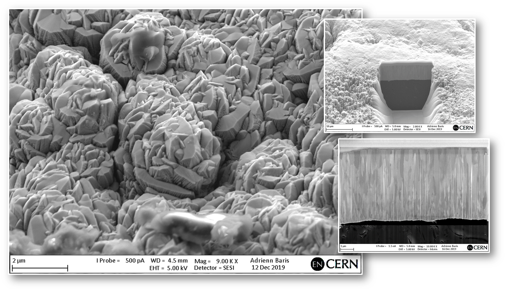
Mo coated MoGr blocks for target collimators
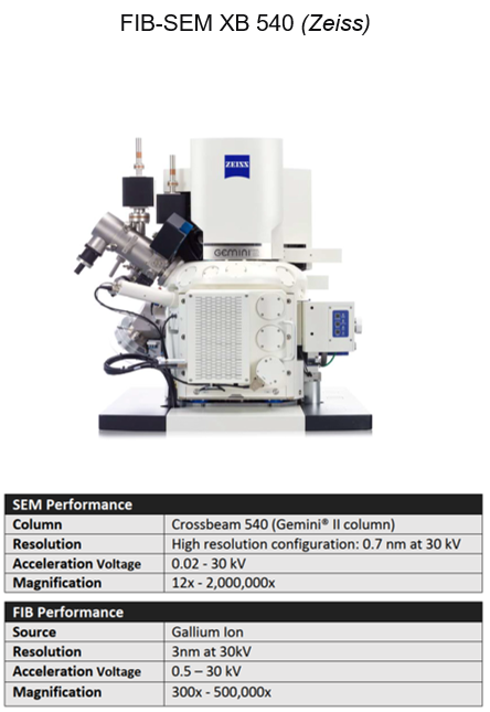
Further information and technical specifications of the equipment can be found here: XB540
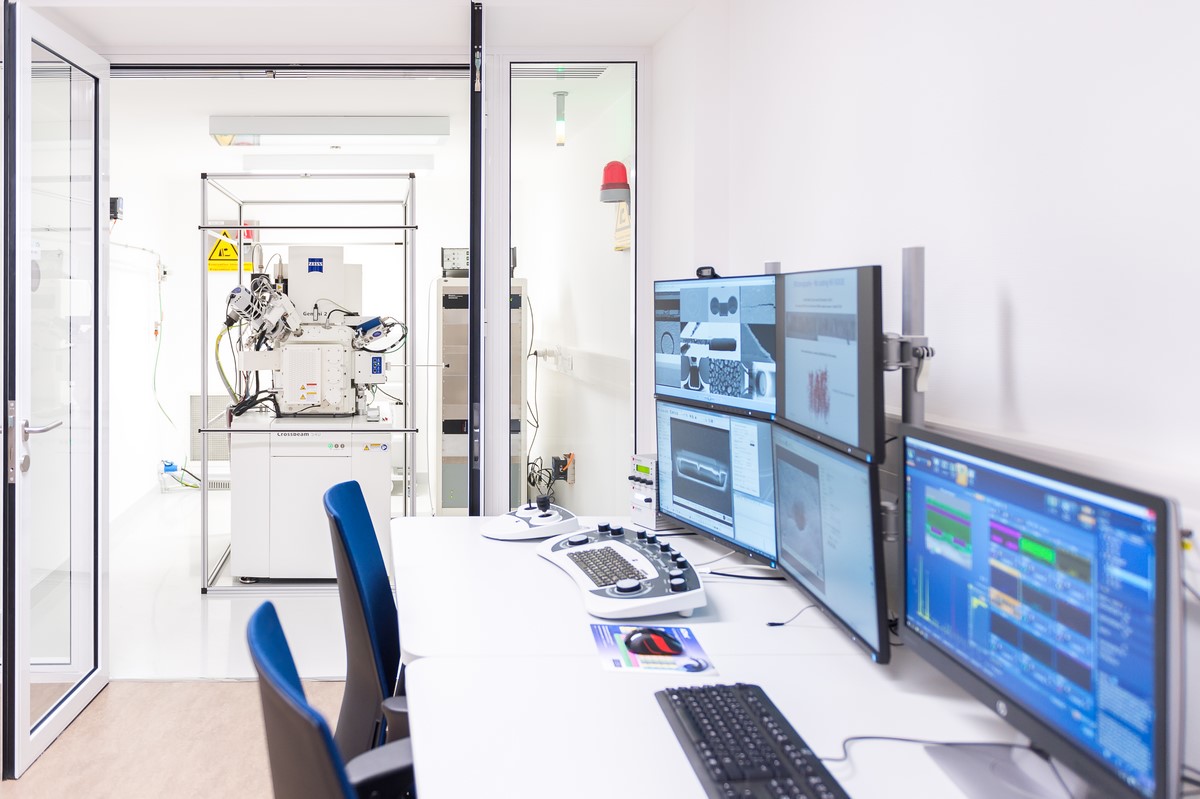
FIB-SEM system in 376/R-014