Focused Ion Beam (FIB) tomography is a highly specialised technique that made possible through the combined use of SEM and FIB (dual beam system). It is considered a semi-destructive technique. The role of the FIB is to make systematic cuts along the specified area of a sample. These areas are traditionally measured in the micro/nanometre range. The SEM then takes sequential images for each cut. This series of images is subsequently transferred into an appropriate software to form a 3D representation of the sample inspected volume.
The software reconstruct the 3D volume by applying different algorithms to segment the image by separating the pixels of the region of interest from the background or other objects in the image. After several computational steps, the 3D model of the features of interest can then be visualised.
This 3D reconstruction can be highly effective for analyses such as:
- Failure analysis including defect imaging
- Microstructure segregation studies
- Inclusion or other features distribution studies
The 3D reconstruction allows the material experts to gain a multi-angle view of the represented sample allowing them to make a more sophisticated appraisal of the material condition. This level of sophistication is improved with the collaborative functions of the EDX software which promotes the use of compositional mapping at each of these SEM slice images. This application can allow for such analyses as:
- Grain structure in 3D (visible grain boundaries in 3D)
- Compositional distribution
- Quantification of composition in 3D
For a fully effective application of this technique, the 3D reconstruction requires an analysis duration of several days.
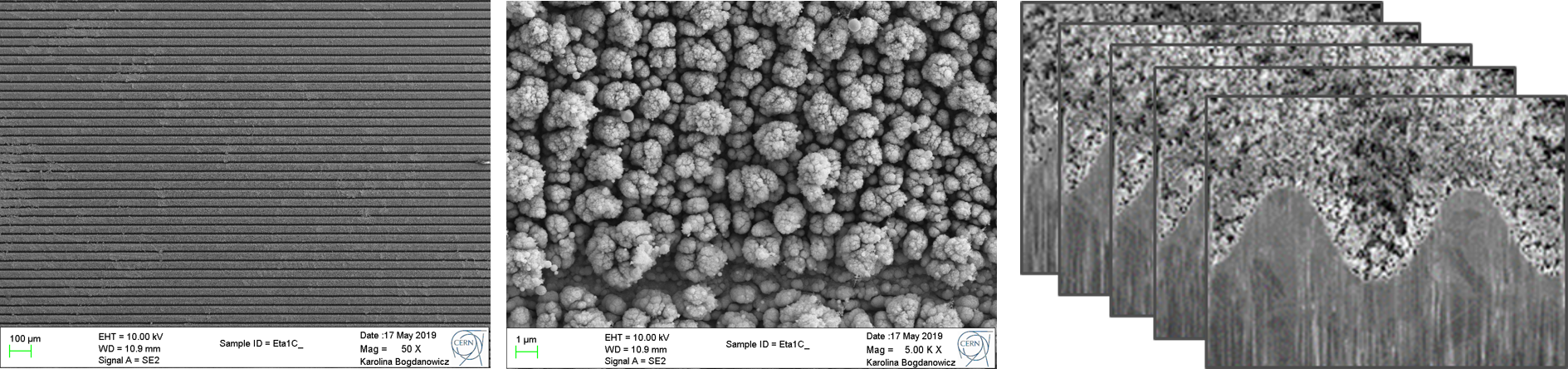
An example of a FIB tomography performed on a Cu-LESS sample is here included.