One of the most crucial aspects of the Focused Ion Beam (FIB) technology is the ability to prepare incredibly thin lamellae specimens which can be used in conjunction with other microscopy techniques such as Scanning Transmission Electron Microscopy (STEM) internally within the FIB chamber.
The use of the FIB allows for the milling of the specimen down to suitable thickness as the analysis of the lamella depends on the transmission of electrons through the sample material. Lamella preparation is a highly specialised technique, one which demands many hours of preparation time. It exist the possibility to characterize the lamella produced in this way internally at the materials laboratory (see below STEM technique details) but also lamellae can be produced for external analysis through Transmission Electron Microscopy (TEM).
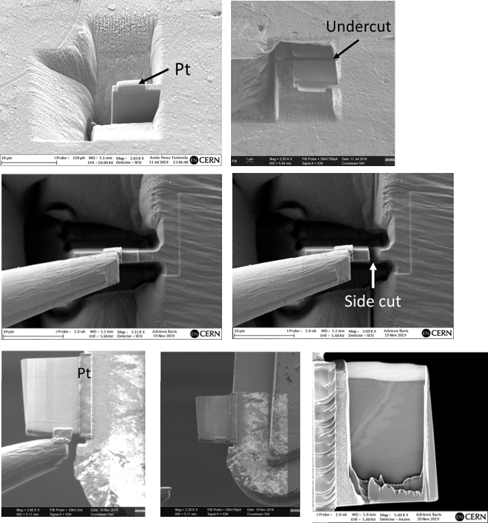
The essential process of this application is simplified in the following image.
Scanning Transmission Electron Microscopy (STEM)
A further technique within the FIB-SEM arsenal is STEM. This technique offers a bridge between Scanning Electron Microscopy (SEM) and TEM.
This technique accommodates for samples with a greatly reduced thickness as the path of the electrons are primarily through transmission allowing the signal to be received below the sample. For this to be possible, the sample must be milled down to a thickness no greater than 100 nm. This thickness of sample is produced through the formation of the lamellae structure mentioned above.
The STEM detector is built up by several segments and allows for the collection of bright field (BF), dark field (DF) and high-angle annular dark field (HAADF) micrographs. The use of the STEM mode of the Cross Beam 540 provides:
- The highest magnification and resolution studies possible with the XB540 machine (0.5 nm)
- Chemical analyses coupled with the EDX technique
- Nanoscale resolution studies of structure and elemental distribution
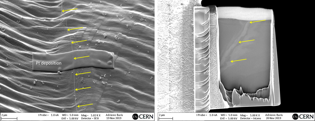
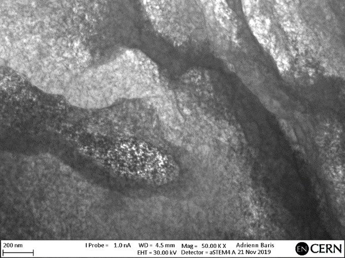 Lamella extraction on deformed Niobium Single Crystals and STEM image of the dislocation substructure in the vicinity of a shear band formed in the crystal during deformation